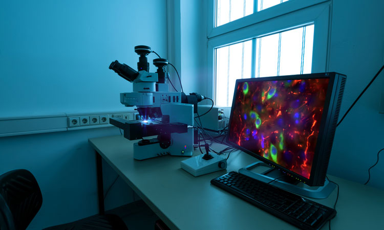
Super-resolution microscopy builds multicolour 3D images from 2D
Posted: 1 October 2018 | Iqra Farooq (Drug Target Review) | No comments yet
Researchers have developed a method of exploiting fluorescence in microscopy to generate 3D images of organelles and structures within cells from 2D images…
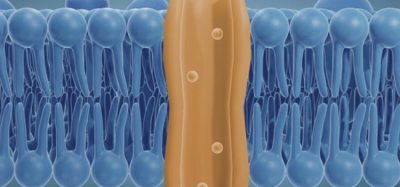
Super-resolution microscopy is a method that can ‘see’ beyond the limits of light. By exploiting fluorescence, super-resolution microscopy allows scientists to observe cells and their interior structures and organelles.
The interior structures of cells are made up of multiple proteins, and and microscopy can typically use only one or two fluorescent colours. As such, it can be difficult to distinguish between different proteins and decipher the complex architecture, to understand the underlying assembly mechanisms of the cell’s interior structures.
Scientists from the Suliana Manley lab at Ecole Polytechnique Federale De Lausanne (EPFL) have developed a technique to analyse and reconstruct super-resolution images and re-align them in a way that multiple proteins can be placed within a single 3D volume.
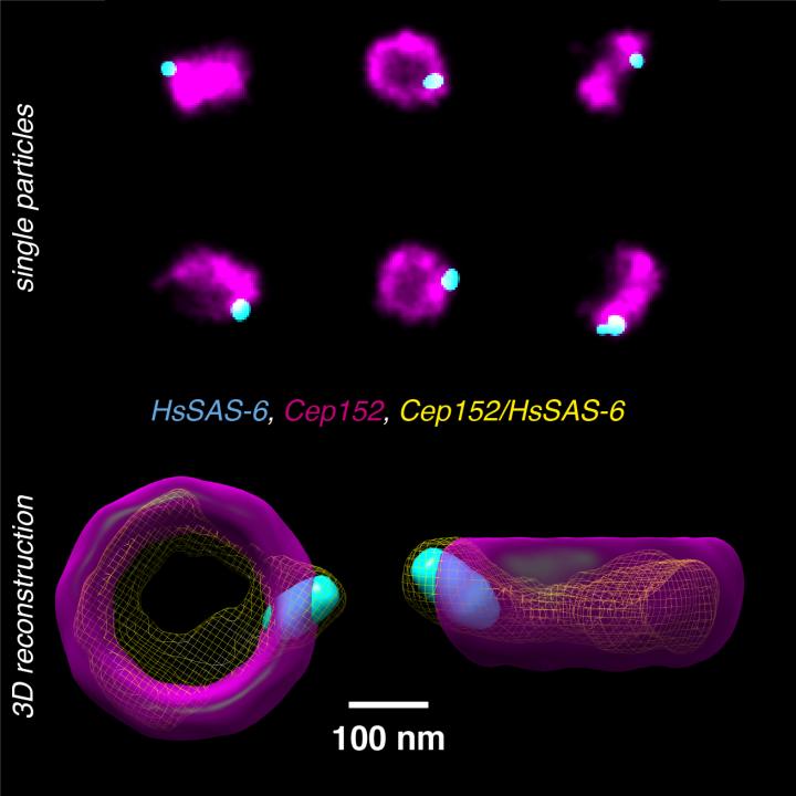
Human centrioles labelled with antibodies against two proteins (Cep152, HsSAS-6) and imaged using super-resolution microscopy. From many individual particles showing projections of the centriole complex in various orientations (upper panel), by using a fused intermediate (yellow, lower panel), the newly developed method allows now to reconstruct a multicolor 3D model (lower panel). Credit: Christian Sieben/EPFL.
Images taken with large field-of-view super-resolution microscopy, where each image contains hundreds of 2D projections of a labelled structure in parallel, can be used and realigned so that multiple proteins can be visualised in a three-dimensional way.
The researchers tested the method on human centriole complexes. Centrioles are pairs of cylindrical molecular assemblies that are crucial in helping cell division. They are positioned near the nucleus and are involved in the development of spindle fibres within the process of cell division.
Using the mutlicolour super-resolution reconstruction method, the researchers uncovered the 3D architecture of four of the proteins that are critical for centriolar assembly during organelle biogenesis.
“With this method, if the proteins in the structure can be labeled, there is no limit to the number of colors in the 3D reconstruction,” said Professor Suliana Manley. “Plus, the reconstruction is independent of the super-resolution method used, so we expect this analysis method and software to be of broad interest.”
Related topics
Disease Research, Drug Discovery Processes, Drug Targets, Imaging, Research & Development, Screening
Related organisations
Ecole Polytechnique Fédérale De Lausanne (EPFL)
Related people
Professor Suliana Manley