


news
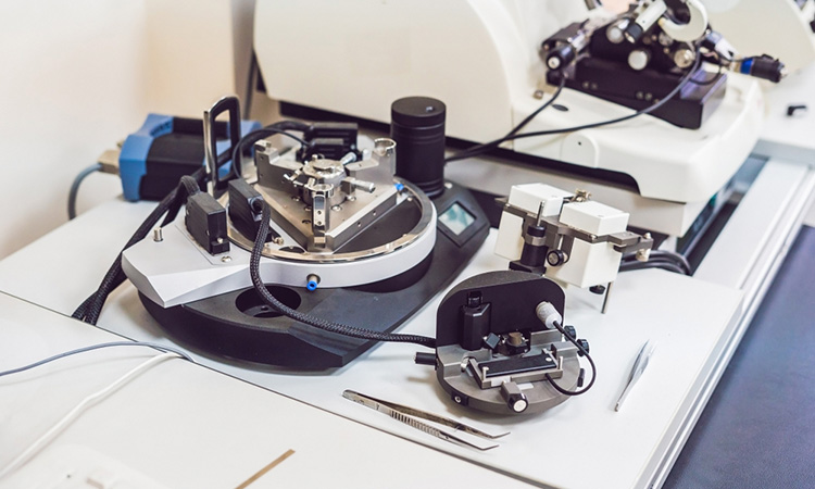
Atomic force microscopy used to study endonuclease digestion of DNA nanostructures
A team of researchers have used microscopy techniques to monitor DNA degradation and anticancer drug release from nanostructures.
List view / Grid view
A team of researchers have used microscopy techniques to monitor DNA degradation and anticancer drug release from nanostructures.

A fluorescence imaging technique has allowed scientists to observe RNA in real time using single-molecule localisation microscopy.
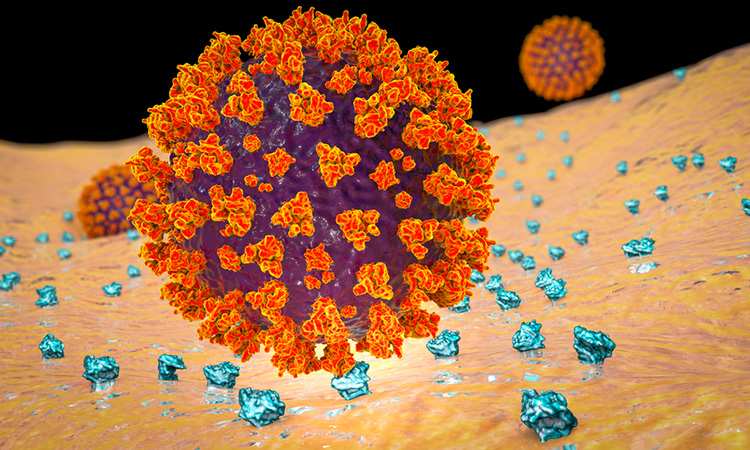
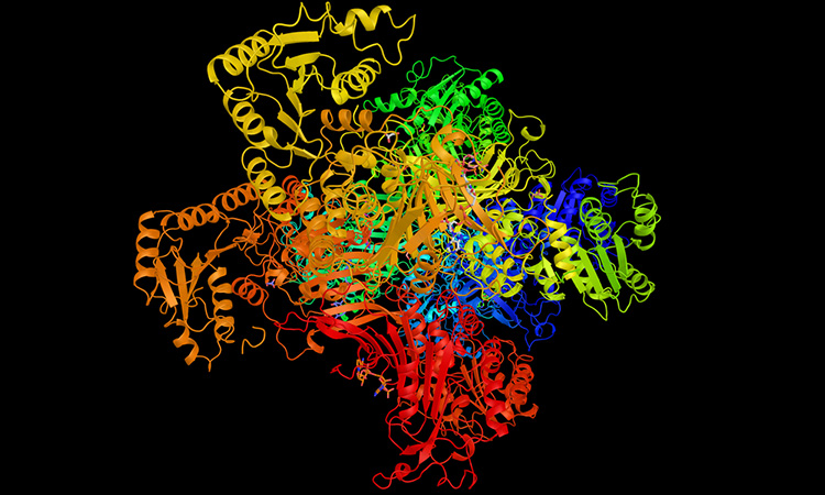
Comparing the original SARS-CoV-2 Spike protein with a mutated version, researchers have potentially revealed why the mutated version is dominant.
21 January 2021 | By Yokogawa Life Innovations
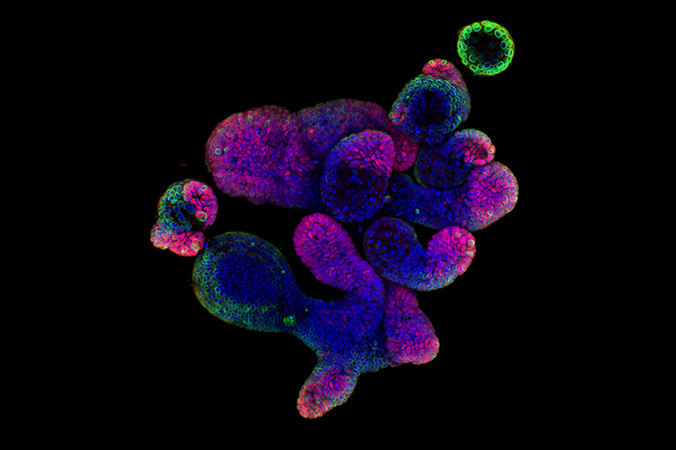
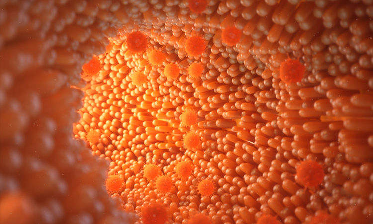
Watch our on-demand webinar and learn how image-based phenotypic screening relies on extraction of multivariate information from cells cultured in a large number of screened conditions. In this webinar, we explore the application of complex and biologically relevant model systems for drug screening, such as small intestinal organoids.
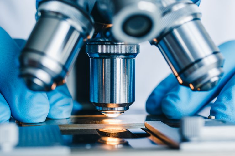
In this article, Drug Target Review’s Hannah Balfour discusses three of the latest developments in imaging for disease research and drug development.
This tissue-specific handbook brings you key publications, in-house protocols and troubleshooting recommendations for your organoid cell culture.
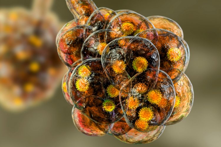
Researchers have revealed how the most severe cases of G6PD deficiency occur, which could help scientists design new drugs for the disease.

Researchers will use the in vitro model to study how respiratory viruses, like SARS-CoV-2, cause Acute Respiratory Distress Syndrome (ARDS) and develop potential interventions.
Using cryo-electron microscopy, researchers have observed the structure of a diarrhoea enteric adenovirus to see how it can survive the stomach.
Researchers have used force atomic microscopy to show the structural dynamics at ribosome stalk proteins when building new proteins.
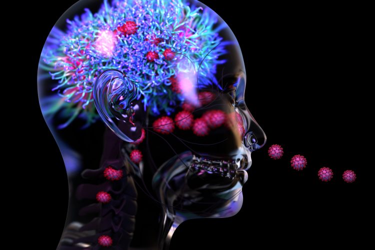
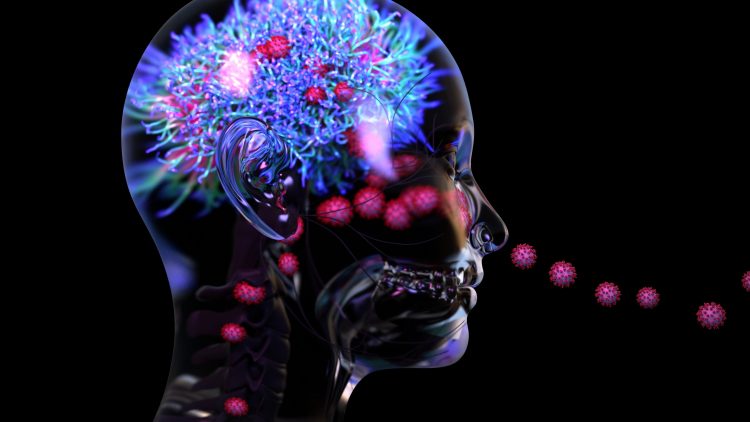
A new study suggests that inflammation and blood vessel damage may be the primary causes of neurological symptoms in COVID-19 patients, instead of the virus infecting the brain.
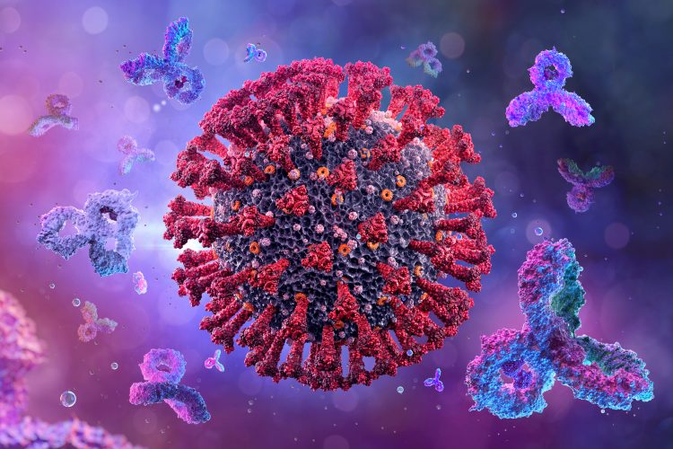
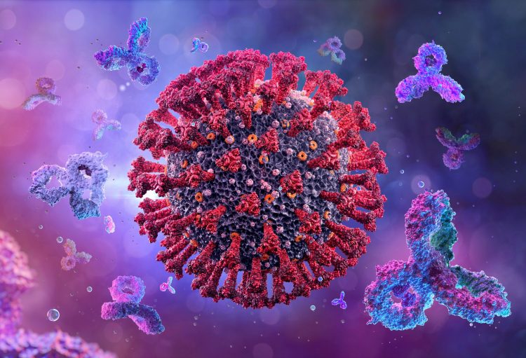
Researchers show that neutralising antibodies targeting the SARS-CoV-2 Spike protein have four distinct structures.
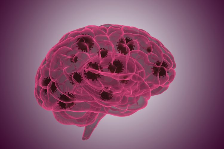
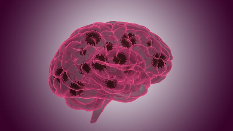
A new study has identified the mechanisms through which the SARS-CoV-2 virus enters the brain and how the immune system responds once it does.
The Vi-CELL BLU automates the widely accepted trypan blue dye exclusion method for cell viability that has historically been performed with a light microscope, pipette, and a hemacytometer.
A new imaging method called FLASH can provide a visualisation of several tissue types in a 3D format, its developers say.