US researchers have uncovered a novel method using a tool used to visualise ion channels in mechanosensory neurons.

A recent study by researchers at Texas Children’s Hospital, Baylor College of Medicine, and Scripps Research Institute, US, has discovered fluorescent dye FM 1-43 as an effective and versatile tool to visualise PIEZO2 ion channel activity in mechanosensory neurons.
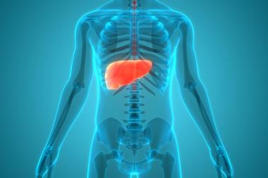
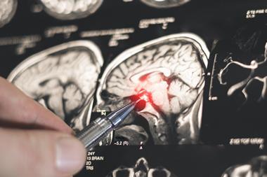
Mechanosensation is the ability to detect dynamic mechanical stimuli like pressure, stretch, and shear stress, and it is crucial for the function of many physiological processes. Sensory neurons gather mechanical stimuli from the environment and internal organs to convey this information to the brain.
The study, published in Neuron, built on work from a 2010 study that led to the discovery of Piezo1 and Piezo2 as essential components of distinct mechanically-activated ion channels. It was demonstrated that the critical role these channels play in sensing and responding to external and internal mechanical stimuli such as touch, proprioception, and bladder stretch sensation. The broad expression of these receptors in sensory neurons suggests it may have yet undiscovered roles in many organ systems.
“There are no known endogenous chemical agonists for PIEZO2, so identifying where PIEZO2 is expressed is an important clue that a specific cell or tissue may detect and respond to mechanical forces,” said lead author Dr Kara Marshall, Assistant Professor at Baylor College of Medicine, and Investigator at the Jan and Dan Duncan Neurological Research Institute at Texas Children’s Hospital.
“However, one of the challenges in understanding mechanosensation is our inability to specifically identify mechanosensory neurons. Unfortunately, antibodies against PIEZO2 or a PIEZO2-GFP knock-in mouse have had limited efficacy for in vivo localization of PIEZO2 – a technical hurdle that the scientists in the field have struggled with for years and one that has slowed down further progress.”
In their quest to find an in vivo visualisation tool for mechanosensory neurons, this research team turned to fluorescent styryl dye FM 1-43 which partitions into lipid membranes and has long been used to visualize intracellular trafficking pathways in various tissues both in vitro and in vivo.
In addition to its well-documented role in cellular trafficking, FM 1-43 has been proposed to enter cells through a separate mechanism: via sensory ion channels. This dye results in vivid cellular labelling via several sensory ion channels in vitro, and was observed to label many sensory neurons in vivo.
To test how much of FM 1-43 labelling was specifically due to Piezo2-expressing mechanosensory neurons in animals, the team generated transgenic mice that lacked Piezo2 in certain tissues during development. To their absolute surprise, they found that in these animals the FM 1-43 labelling was significantly reduced, suggesting that most labelling in the sensory nervous system depended on Piezo2 activity and was not a result of non-specific affinity for many ion channels, as believed previously.
Based on these and other experiments, the authors concluded that although FM 1-43 is non-specific in vitro, it specifically labels Piezo2-dependent mechanosensory neurons in vivo. The mechanisms underlying these differences in FM 1-43 labeling remain to be explored.