iPS-cell-derived microglia in brain organoids have enabled scientists to understand early brain development and microglia-associated disease.
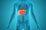
The crucial role of microglia, the brain’s immune cells, in early human brain development, has been uncovered by an international team of researchers. To observe how microglia influence brain cell growth and development, scientists at A*STAR’s Singapore Immunology Network (SIgN) grew brain organoids in the laboratory that mimicked the complex environment of the developing human brain. This has considerable potential for understanding brain maturation and disorders.
Unique pathway
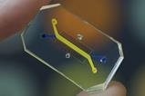
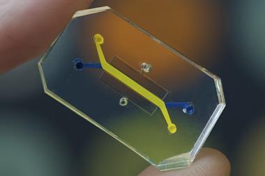
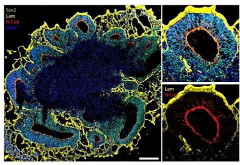
Previous brain organoids have lacked microglia, a key component of early brain development. Therefore, the scientists made a unique protocol to introduce microglia-like cells produced from human stem cells, which were also used to form the brain organoids. The introduced cells behaved like real microglia and influenced other brain cell development within the organoids.
A unique pathway through which microglia interact with other brain cells was discovered, with the researchers finding that microglia have an important role in regulating cholesterol levels in the brain. The microglia-like cells had lipid droplets containing cholesterol which were released and taken up by other developing brain cells in the organoids. The cholesterol exchange was shown to greatly enhance the growth and development of these brain cells, particularly their progenitors.
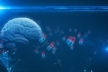
Cholesterol is abundant in the brain and is crucial for neuron structure and function. Neurological disorders, such as Alzheimer's disease and Parkinson’s disease, have been linked to abnormal cholesterol levels.
Proteomics and lipidomics
Led by Dr Markus Wenk, scientists from the Department of Biochemistry at the Yong Loo Lin School of Medicine (NUS Medicine), carried out data acquisition, particularly in the field of lipidomics, to gain insight into the lipid composition and dynamics within the brain organoids containing microglia.
This information was used by another team led by Associate Professor Veronique Angeli from the Department of Microbiology and Immunology at NUS Medicine. They discovered that cholesterol impacts the growth and development of young brain cells in the human brain models. A specific protein is used by microglia to release cholesterol, but when this process of blocked, it initiates organoid cell growth, resulting in larger brain models.
“Our next focus will be finding out how we can regulate cholesterol release to optimise brain development and slow down, or prevent, the onset of neurological conditions.”
Dr Angeli noted that this finding is particularly impactful as “we finally understand how cholesterol is transported.” She continued: “Our next focus will be finding out how we can regulate cholesterol release to optimise brain development and slow down, or prevent, the onset of neurological conditions.”
Dr Olivier Cexus from the University of Surrey and previously from A*STAR, used proteomic and lipidomic analysis to progressively decipher the complex molecular interactions in the brain organoids. This provided a deeper understanding into the metabolic cross-talks involved in brain development and potential implications for diseases.
Also, quantitative proteomics was used by Dr Radoslaw Sobota at A*STAR's Institute of Molecular and Cell Biology (IMCB) and his team at the SingMass National Laboratory for Mass Spectrometry to uncover changes in protein. Their analysis of the protein composition of the organoids further confirmed the study’s discovery.
Potential therapies
Dr Jerry Chan, co-author of the study and Senior Consultant at the Department of Reproductive Medicine, KK Women’s and Children’s Hospital, and Senior National Medical Research Council Clinician Scientist, explained: “There is currently a lack of tools to study how microglia interacts with the developing brain. This has hampered the understanding of microglia-associated diseases that play an important role during the early development of conditions such as autism, schizophrenia, and neurodegenerative diseases such as Alzheimer’s and Parkinson’s disease.
He continued: “The development of these novel microglia-associated brain organoids with same-donor pluripotent stem cells gives us an opportunity to study the complex interactions between microglia and neurons during early brain development. Consequentially, this may enable us to study the role of microglia in the setting of diseases and suggest ways to develop new therapies in time.”
The study was published in Nature.