Norovirus structure imaged, providing potential for vaccine development
Posted: 17 June 2019 | Victoria Rees (Drug Target Review) | No comments yet
Researchers have been able to image noroviruses, providing significant information for the development of therapeutics.
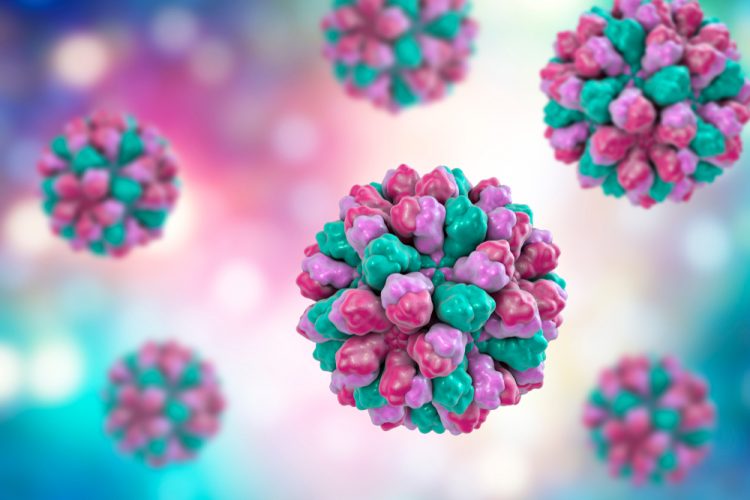
A new study has used high-resolution imaging to identify the outer layer, or capsid, of noroviruses. The findings could lead to vaccine developments to prevent outbreaks.
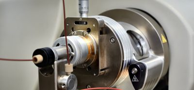
Researchers at Cold Spring Harbor Laboratory (CSHL), New York, used high-resolution cyro-electron microscopy to identify the structure of the virus shells.
Noroviruses are a leading cause of food-borne illness outbreaks, accounting for 58 percent of cases. A total of 685 million cases are reported each year.
“Previously, it was thought that the norovirus shells exist in single-sized assemblies consisting of 180 building blocks and 90 surface spikes. What we found was an unexpected mixture of different shell sizes and shapes. We found a smaller form, which consists of just 60 building blocks with 30 surface spikes placed further apart. We also found larger shells made out of 240 building blocks with 120 surface spikes that are lifted significantly above the base of the shell and form a two-layered architecture that could interact differently with the human cells,” said James Jung, from CSHL.
The team observed that the distance and orientation of the spikes varied across the different strains of noroviruses. They say this means each strain will interact differently with human host cells, so the way that antibodies bind will be different. As such, vaccines can be formulated to take into account the variations across strains and structural forms.
The findings were published in PNAS.
Related topics
Drug Targets, Imaging, Microscopy, Structural Biology, Vaccine
Related conditions
norovirus
Related organisations
Cold Spring Harbor Laboratory, PNAS
Related people
James Jung