Researchers use imaging to reveal early stages of colon cancer
Posted: 2 December 2019 | Victoria Rees (Drug Target Review) | No comments yet
A study has demonstrated how mutations in early colon cancer prevail and grow into malignancies, using fluorescent imaging.

Researchers have revealed how stem cell mutations arise and spread throughout a widening field of the colon until they eventually become a malignancy. According to the team, their technique could lead to therapeutic developments and earlier treatment of the disease.
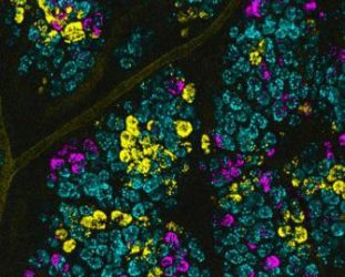
High magnification image of fluorescent intestinal stem cells. Each fluorescent colour is used as a barcode to visualise human colon cancer-causing mutations in mice (credit: Duke Health).
The study was conducted at the Duke Cancer Institute at Duke University, US.
Using an innovative modelling system in mice, the researchers visually tagged colon cancer mutations by causing stem cells to grow. This allowed them to identify the prevailing mutations, which could be visualised in the animals.
Biomarkers are redefining how precision therapies are discovered, validated and delivered.
This exclusive expert-led report reveals how leading teams are using biomarker science to drive faster insights, cleaner data and more targeted treatments – from discovery to diagnostics.
Inside the report:
- How leading organisations are reshaping strategy with biomarker-led approaches
- Better tools for real-time decision-making – turning complex data into faster insights
- Global standardisation and assay sensitivity – what it takes to scale across networks
Discover how biomarker science is addressing the biggest hurdles in drug discovery, translational research and precision medicine – access your free copy today
The team tagged several common colon cancer mutations in the stem cells of a single tumour to create a fluorescent barcode. When transferred to a mouse, the colours of the stem cells could be tracked, revealing the cellular and molecular dynamics of the pre-cancerous events.
The researchers suggest that their technique can be used to discover field cancerisation, which is suggested to be the defining event that initiates the process of cancer growth.
“This study provides new insight into the previously invisible process in which mutant pre-cancerous stem cells spread throughout the colon and seed cancer,” said Dr Joshua Snyder, Assistant Professor in the Departments of Surgery and Cell Biology at Duke and corresponding and co-senior author.
“Our technique sets a firm foundation for testing new therapies that interrupt this early, pre-malignant process. We hope to one day target and eliminate these stealth precancerous cells to prevent cancer,” Snyder continued.
The findings were published in Nature Communications.
Related topics
Disease Research, Drug Targets, Imaging, Oncology, Research & Development, Stem Cells
Related conditions
Colon cancer
Related organisations
Duke Cancer Institute at Duke University
Related people
Dr Joshua Snyder