Imaging technique allows study of early stage Alzheimer’s disease
Posted: 26 March 2020 | Hannah Balfour (Drug Target Review) | No comments yet
Scientists used optical photothermal spectroscopy (O-PTIR) to image murine neurons affected by early stage Alzheimer’s disease, providing insight into the progression of the disease.
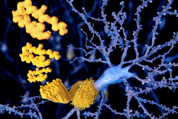
Neuron (purple) with beta-amyloid plaque forming (yellow).
In order to study the early stages of Alzheimer’s disease (AD), researchers have used a new imaging method, called optical photothermal spectroscopy (O-PTIR), to visualise the protein structures within live nerve cells.
According to researchers, one of the major roadblocks to effective AD therapies is that they have been unable to study the early protein structural changes that occur before visible plaques appear. Essentially, they have been unable to answer: what happens in the nerve cells before amyloid plaques appear and why do nerve cells die?
“This is a question that researchers have long struggled to find answers to. We have not had enough imaging techniques to study the structural changes in nerve cells. This is required to detect very early changes, and therefore potentially understand the triggers,” said researcher Oxana Klementieva, group leader for medical microscopy at Lund University, Sweden.
Biomarkers aren’t just supporting drug discovery – they’re driving it
FREE market report
From smarter trials to faster insights, this report unpacks the science, strategy and real-world impact behind the next generation of precision therapies.
What you’ll unlock:
- How biomarkers are guiding dose selection and early efficacy decisions in complex trials
- Why multi-omics, liquid biopsy and digital tools are redefining the discovery process
- What makes lab data regulatory-ready and why alignment matters from day one
Explore how biomarkers are shaping early drug development
Access the full report – it’s free!
In a study published in Advanced Science, Klementieva and her team used O-PTIR to study protein structures within nerve cells affected by early-stage AD in mice, before cell death occurred. The process did not require the use of chemical processing of nerve cells, unlike other imaging techniques, which can affect the structure of neurons.
“We saw that the structure of the protein changes in different ways depending on where in the nerve cell it is. So far, there have been no methods that can produce these types of images, giving us insight into what the first molecular changes in neurons actually look like in Alzheimer’s disease,” explained Klementieva.
In previous research projects, Klementieva and colleagues demonstrated that early structural changes of beta-amyloid happen before the AD hallmark amyloid plaques appear. They now intend to investigate where in the cell these structural changes occur in the hopes it may explain the mechanisms underlying AD.
“If it depends on several different mechanisms, something I believe to be the case, we would need different types of treatments,” said Klementieva.
The team believe the O-PTIR technology could also be leveraged in studies of related diseases like Parkinson’s, Lewy Body dementia and frontal lobe dementia.
Related topics
Disease Research, Drug Targets, Imaging, Neurons, Neurosciences, Protein, Proteomics, Technology
Related conditions
Alzheimer’s disease, Dementia, frontal lobe dementia, Lewy body dementia, Parkinson's disease (PD)
Related organisations
Lund University
Related people
Oxana Klementieva