

Researchers capture images of cells infected with SARS-CoV-2
Posted: 11 September 2020 | Victoria Rees (Drug Target Review) | 1 comment
Using scanning electron microscopy, researchers have captured images of epithelial cell cultures infected by SARS-CoV-2.
A group of researchers has announced that they have produced images of the infectious form of SARS-CoV-2, the virus that is causing the COVID-19 pandemic. The images were taken in epithelial cell respiratory tract cultures.
The images were taken by a team from the University of North Carolina (UNC) School of Medicine, US, laboratory of Assistant Professor Camille Ehre, in collaboration with both the labs of Dr Ralph Baric and and Dr Richard Boucher.
The researchers say they captured these graphic and easily understood images to illustrate how intense the SARS-CoV-2 infection of the airways can be.
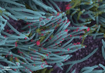
In a laboratory setting, the team inoculated the SARS-Co-V-2 virus into human bronchial epithelial cells, which were then examined 96 hours later using scanning electron microscopy.
A higher power magnification image shows the structure and density of SARS-CoV-2 virions (red) produced by human airway epithelia [credit: Ehre Lab, UNC School of Medicine].
The images, re-colorised by UNC medical student Cameron Morrison, show infected ciliated cells with strands of mucus (yellow) attached to cilia tips (blue). Cilia are the hair-like structures on the surface of airway epithelial cells that transport mucus (and trapped viruses) from the lung. A higher power magnification image shows the structure and density of SARS-CoV-2 virions (red) produced by human airway epithelia. Virions are the complete, infectious form of the virus released onto respiratory surfaces by infected host cells.
According to the researchers, this imaging research helps to illustrate the incredibly high number of virions produced and released per cell inside the human respiratory system. The large viral burden is a source for spread of infection to multiple organs of an infected individual and likely mediates the high frequency of COVID-19 transmission to others.
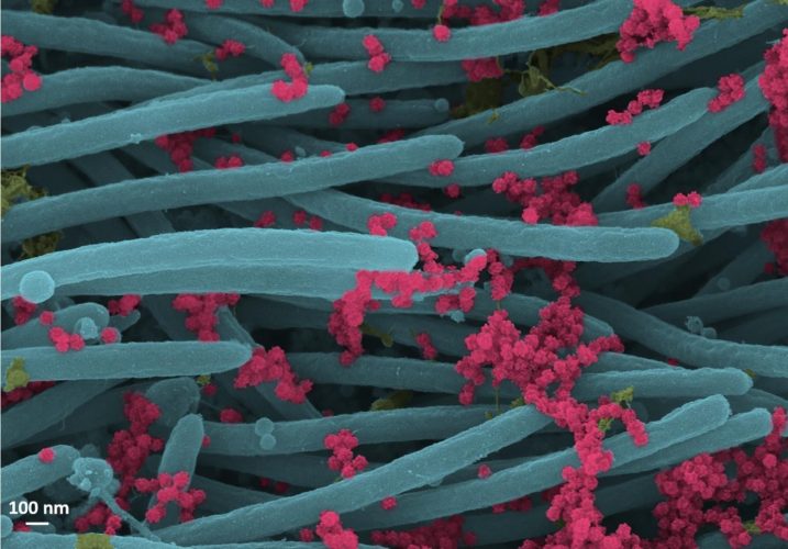
SARS-CoV-2 virions (red) [credit: Ehre Lab, UNC School of Medicine].
The team say these images make a strong case for the use of masks by infected and uninfected individuals to limit SARS-CoV-2 transmission.
The images were published in the New England Journal of Medicine.
Related topics
Disease Research, Imaging, Microscopy, Research & Development, Targets
Related conditions
Covid-19
Related organisations
University of North Carolina (UNC)
Related people
Assistant Professor Camille Ehre, Dr Ralph Baric, Dr Richard Boucher


I would like to know the magnification of the “A higher power magnification image shows the structure and density of SARS-CoV-2 virions (red) produced by human airway epithelia [credit: Ehre Lab, UNC School of Medicine].”
Knowing the magnification would help in identifying the relationship of the lung cilia size to the sample size.
Thank you