

whitepaper
Article: Defeating COVID-19: The science behind a new ELISA
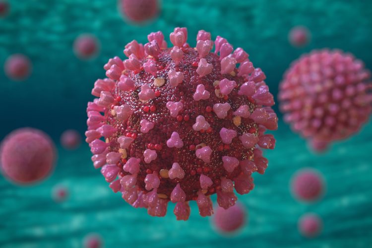
Discover how a new ELISA against S1-RBD for COVID-19 seroconversion detection can accelerate discovery to facilitate vaccine breakthroughs.
List view / Grid view
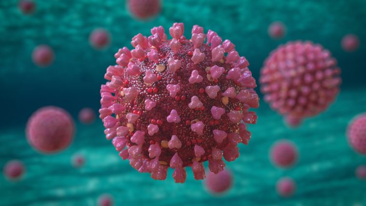
Discover how a new ELISA against S1-RBD for COVID-19 seroconversion detection can accelerate discovery to facilitate vaccine breakthroughs.
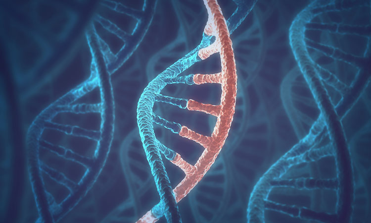
Researchers have elucidated the 3D structure of the Taspase 1 enzyme, known to be involved in a range of cancers.
Researchers have created a single-celled synthetic organism able to grow and divide that could be used to produce drugs and detect disease.
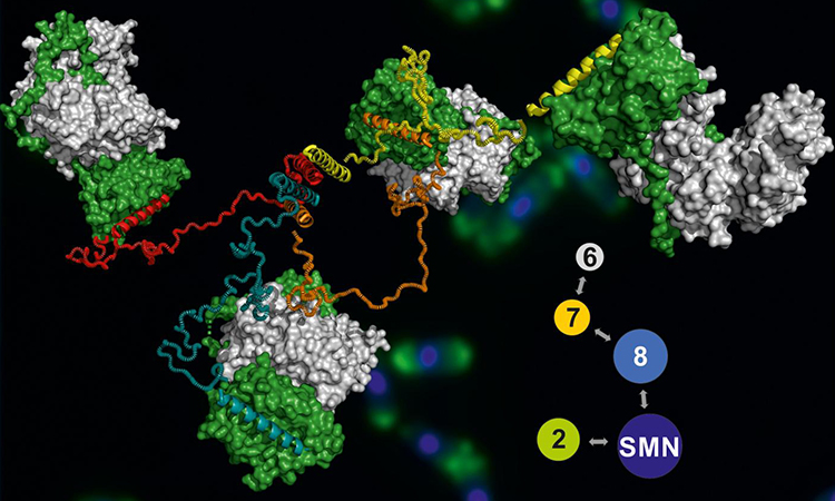
Researchers have imaged the entire Survival Motor Neuron complex using X-ray diffraction analysis, among other techniques.

Working with RNA requires rigorous nuclease contamination controls in place. Stock up on Nuclease-Free tubes tips and buffers #Back2Lab
Using cryo-electron microscopy, researchers have imaged how the SARS-CoV-2 Spike protein changes with the D614G mutation to enable faster spread of infection.
A large number of nanorobots in the bladders of mice have been visualised to reveal their movement and behaviour.
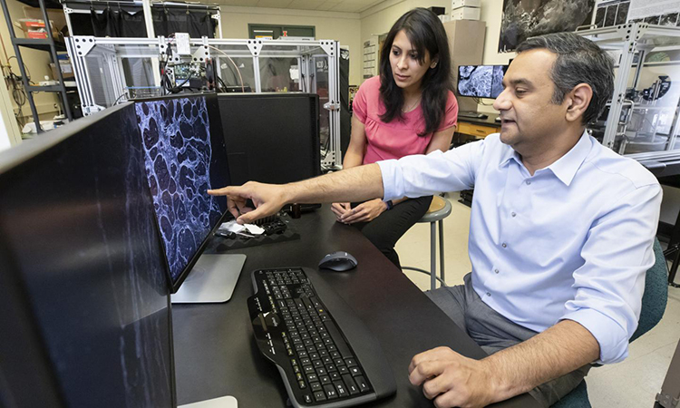
An artificial intelligence platform has been created to enable tens of thousands of microscopy images to be generated in an hour.
Researchers have produced the first 3D image of the Mediator-bound pre-initiation complex, key in the regulation of gene expression.
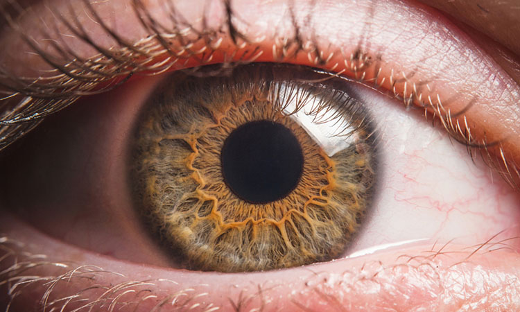
A new imaging technique for photoreceptors has been developed by selectively blocking light used to observe the eye.

New podcast on SARS-CoV-2 molecular testing: From human sample analysis to Wastewater Surveillance.

RiboMinus Bacteria 2.0 Transcriptome Isolation Kits perform efficient transcriptome enrichment from total bacterial RNA. Improve your RNA extraction from bacteria today.

See our guide on DNA purification methods. From organic extraction through automated high-throughput runs, these research solutions make sample preparation easy.
A spectroscopic microscope has been developed by researchers to gather data on biological conformations faster and more accurately.
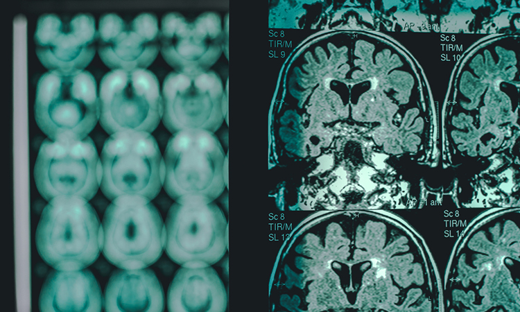
Using X-ray crystallography and cryo-electron microscopy, researchers have elucidated the structure of the SARM1 protein, a target for neurodegeneration.