Muscle cell atlas developed to aid muscular disease and injury research
Posted: 11 March 2020 | Hannah Balfour (Drug Target Review) | No comments yet
Researchers have created a new technical resource atlas which maps the 15 distinct cell types involved in muscle repair for disease and therapy research.
Scientists have created a cell atlas which catalogues the activity of almost every kind of cell involved in muscle repair. The researchers hope their resource could be used to simplify muscular disease research and potentially lead to better injury rehabilitation strategies.
The collaborative efforts of a team from Cornell University’s Meinig School of Biomedical Engineering and Biotechnology Resource Center, both US, allowed researchers to analyse the gene expression signatures of thousands of cells taken from actively regenerating murine muscle tissue.
The paper published in Cell Reports is the culmination of data from approximately 35,000 individual cells. This atlas is a list of the fifteen unique cell types involved in muscle cell repair, itemising their similarities and differences to provide a technical resource for anyone studying skeletal muscle.
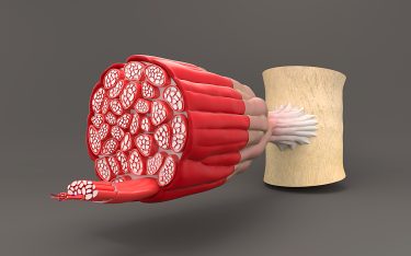
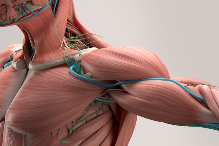
The internal structure of muscles is made up of multiple cell types grouped into fibres.
“Because we have such a large dataset, it helps us frame a number of hypothesis-driven questions about not only which cells are involved, but how they are communicating with each other,” said research leader Ben Cosgrove, assistant professor in the Meinig School of Biomedical Engineering. “This resource enabled us to ask: ‘What molecular signals are one type of cells sending to the other cells within the process of muscle repair?'”
According to the researchers, the muscle cell atlas emphasises how much heterogeneity exists within each cell type, which enables scientists to explore specific molecular variations that may have previously been overlooked.
The team have already used their atlas to identify how proteins called syndecans enable muscle stem cells to decide between replenishing the stem cell population or differentiating into mature myofiber cells that replace damaged muscle tissue during muscle cell repair.
“We took this huge atlas and partitioned it down to the stem cells. And then we organised the stem cells in a way that gives us a framework to think about variation and the choices the cells are making,” Cosgrove said.
They identified that syndecan-related variations may direct how muscle stem cells respond to signals from their neighbouring cells and therefore how they differentiate.
“Now that we know more about what is happening in the healthy, normal adult repair process, we can ask ‘How do the cellular players get misallocated and misactivated in the disease settings?'” Cosgrove said. The team is currently using the atlas to study muscle repair deficiency in ageing and muscular dystrophy.
Related topics
Analysis, Analytical Techniques, Disease Research, Drug Targets, Genomics, Informatics, Protein, Protein Expression, Proteomics, Regenerative Medicine, Sequencing, Targets
Related conditions
Muscle degeneration, Muscular dystrophy
Related organisations
Cornell University’s Biotechnology Resource Center, Cornell University’s Meinig School of Biomedical Engineering
Related people
Ben Cosgrove