New precise method can visualise individual blood cells in the brain
Posted: 31 August 2021 | Anna Begley (Drug Target Review) | No comments yet
Researchers have developed an inexpensive method for visualising blood flow in the brain that can discern the motions of individual blood cells.
A team from the Skolkovo Institute of Science and Technology and Saratov State University, both Russia, have created an inexpensive method for visualising blood flow in the brain. The technique, which integrates optical microscopy and image processing, is so precise that it can detect the motions of individual red blood cells without the use of dyeing agents or genetic engineering.
“Our method uses what is known as frame-by-frame filtering to process brain images obtained with an ordinary optical microscope available at any lab,” explained lead author Maxim Kurochkin. “It allows us to discern single moving red blood cells and build a highly detailed map of the vasculature, down to the smallest capillaries. This in turn makes it possible to accurately assess blood flow rates in the vessels via a technique called particle image velocimetry.”
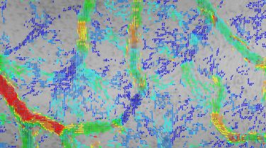
Red blood cell velocity distribution measured and mapped via the new method devised by the Skoltech-SSU team. Each arrowhead corresponds to one cell, with the velocity colour-coded from blue (slow) through green (moderate) to red (fast) [credit: Maxim Kurochkin/Skoltech].
There are already visualising methods, although they can be inaccurate and expensive. One highly precise technique involves injecting fluorescent dyes into the blood flow and detecting the infrared light they emit, but dyes are toxic and also may distort mapping results by affecting the vessels. Alternatively, researchers employ genetically modified animals, whose interior lining of blood vessels is engineered to give off light with no foreign substances involved. However, both methods are very expensive.
The team demonstrated the method’s applicability using a mouse brain and a chicken embryo. They began by using the vascular networks of the chicken embryo to demonstrate the possibility of mapping even the tiniest capillaries, in which red blood cell motions may be inconstant.
The researchers then tested the method on a rat brain vasculature. The technique proved capable of mapping blood vessel networks even for a system with vessels that are harder to reach, with no individual red blood cells discernible, only the colour patterns associated with groups of vessels.
The team hope that their method will lead to a better comprehension of the physiology of endothelial cells which could pave the way to more advanced understandings of coronary heat diseases, as well as new therapies. “Understanding the behaviour of objects that wind up in the blood flow is not limited to the naturally occurring ruptured atherosclerotic plaques,” Kurochkin continued. “In targeted drug delivery, artificial microcapsules with therapeutic agents may be introduced into the bloodstream and vasculature models are indispensable for predicting exactly what happens to them there.”
The study was published in The European Physical Journal Plus.
Related topics
Analytical Techniques, Drug Delivery, Imaging, In Vivo, Label-Free, Microscopy
Related conditions
Cardiovascular disease
Related organisations
Saratov State University, Skolkovo Institute of Science and Technology
Related people
Maxim Kurochkin