


whitepaper
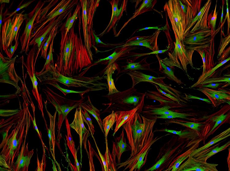
Guide: Five ways biophysical tools help brain studies
Learn why researchers turned to biophysical methods to expose the molecular mechanisms of neurodegenerative diseases.
List view / Grid view
Learn why researchers turned to biophysical methods to expose the molecular mechanisms of neurodegenerative diseases.

Learn practical tips from eight Principal Investigators about how to start your own lab.
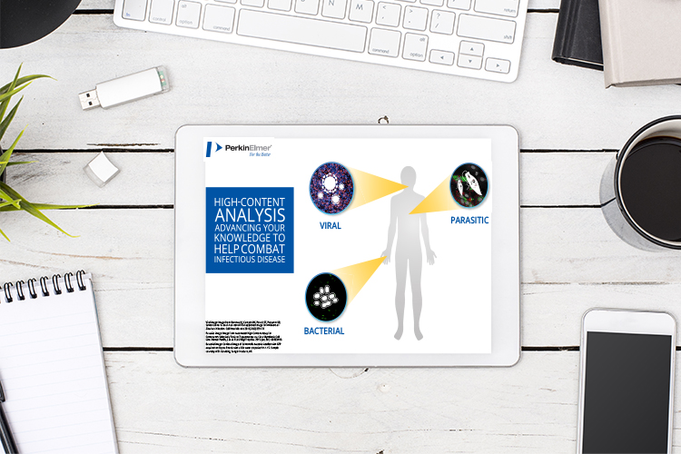
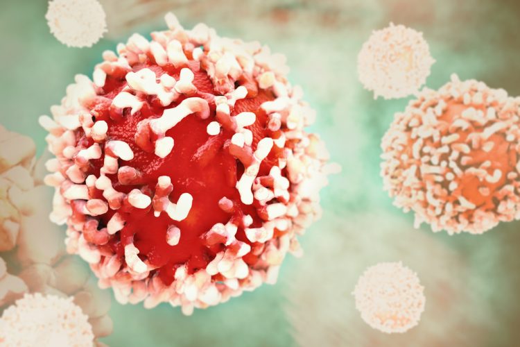
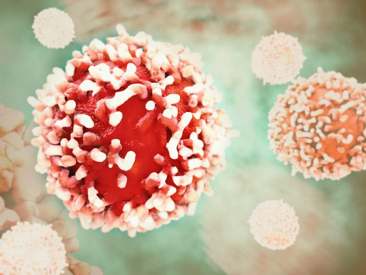
Learn how you could use high-content analysis for functional & phenotypic assays in your infectious disease research or drug discovery.
A group of researchers has used cryo-EM to discover the structure of the remdesivir-bound RNA complex of SARS-CoV-2 and explain how the drug inhibits COVID-19 viral replication.
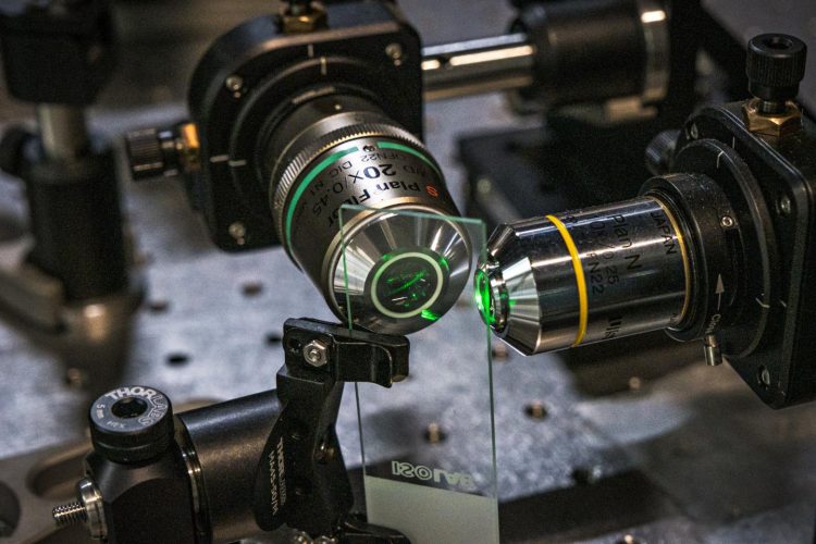
The developers of the ultra-precise single-molecule microscope demonstrated it can resolve interactions between molecules within living cells and is compatible with existing microscopes.

By combining quantitative phase microscopy and molecular vibrational imaging, researchers have created a new label-free microscopy technique.
Using cyro-electron microscopy, researchers have imaged the binding site between a molecule and the tumour suppressor protein PP2A, enabling optimisation of the drug compound.
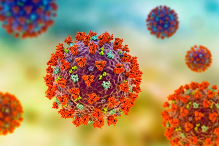
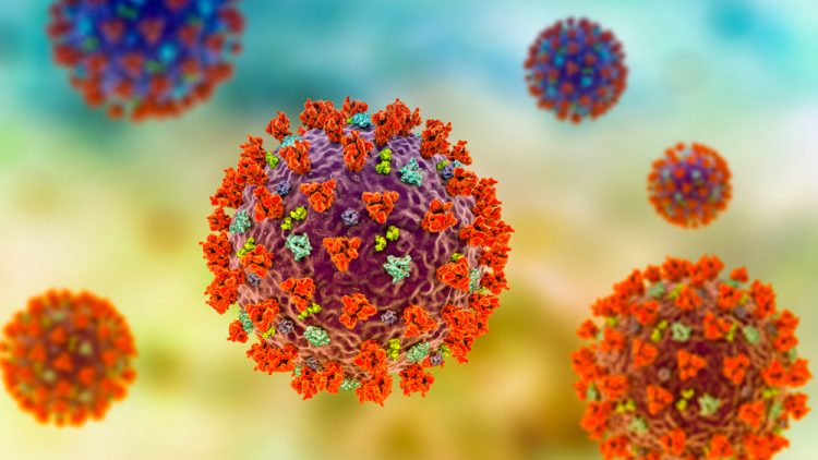
Researchers in the UK have selected nanobodies that bind with high affinity to the Spike protein on the COVID-19 coronavirus, enabling stabilisation for imaging.
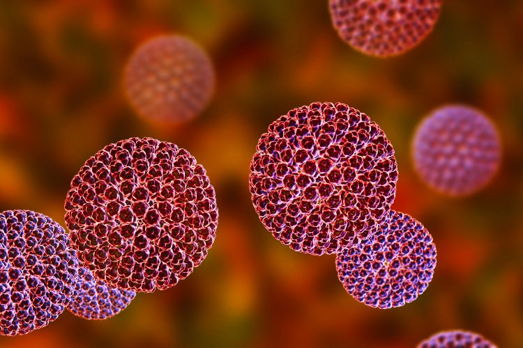
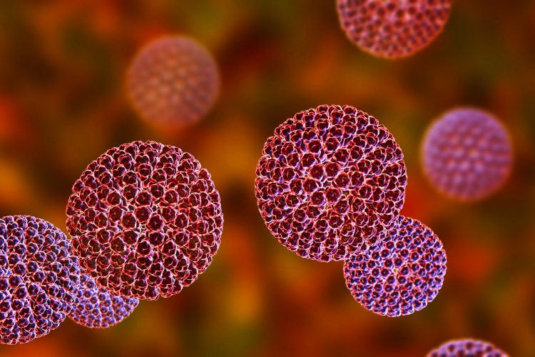
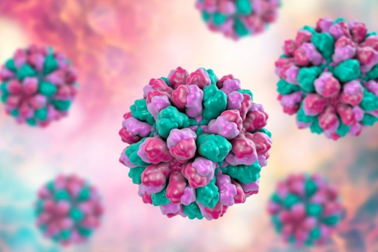
Researchers hope that by revealing the rotavirus VP3 protein structure and mRNA capping functions, novel antivirals could be designed to prevent or combat rotavirus infections.

A new technique called Coded Light-sheet Array Microscopy (CLAM) has been developed by researchers to improve 3D imaging of living specimens.
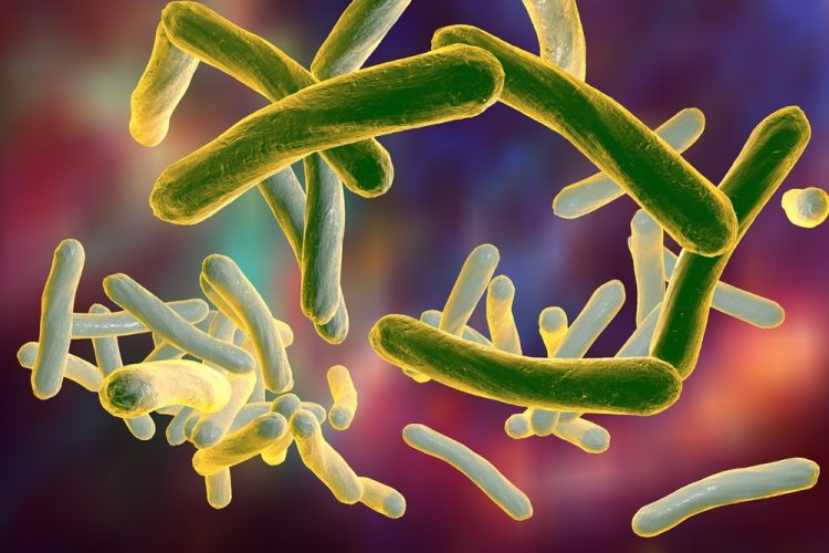

Cryogenic electron microscopy revealed that the vitamin B12 transporter on Mycobacterium tuberculosis acts like a non-selective sluice, transporting both the vitamin and antibiotics.
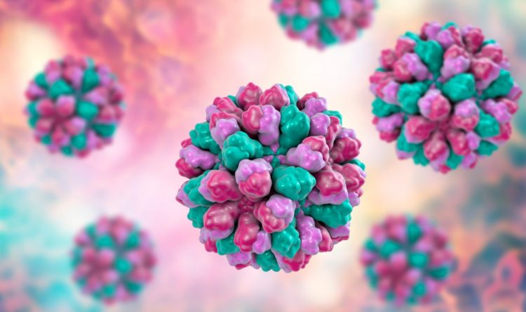
Researchers captured 13,000 images of a mouse norovirus using an electron microscope and compiled the images to reveal the structure of the virus.
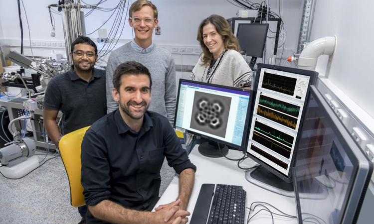
Using AI and deep learning, researchers have enhanced Scanning Probe Microscopy (SPM) and made their automated resource available for scientists.
Scientists have imaged the ball-and-chain mechanism using cryogenic electron microscopy and hope their work could be applied in the design of novel therapeutics.
Researchers have created a new cryogenic electron microscopy (cryo-EM) technique by utilising low-energy electrons in a holographic method.