

news
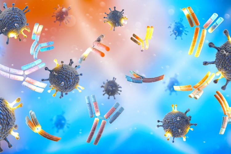
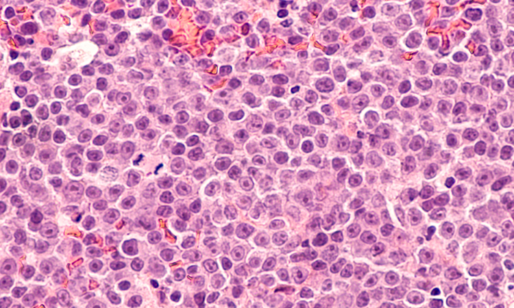
Experimental mAbs show promise against Epstein-Barr virus
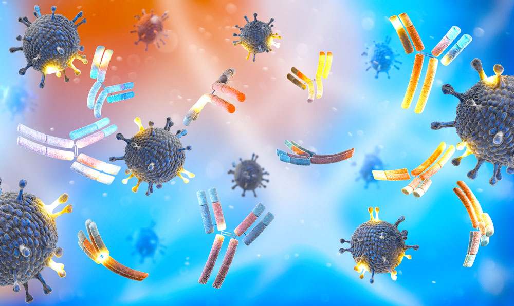
Researchers find monoclonal antibodies provided nearly complete protection against EBV infection and lymphoma when tested in mice.
List view / Grid view
Researchers find monoclonal antibodies provided nearly complete protection against EBV infection and lymphoma when tested in mice.

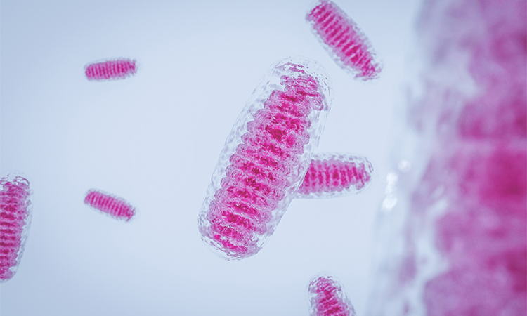
H84T-BanLec has viral-blocking abilities by binding to polysaccharides that are present on the surface of the viruses.
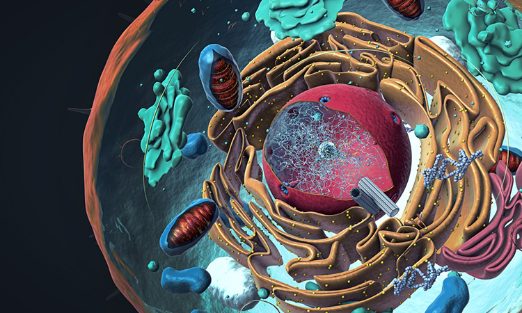
Using cryo-EM, the researchers found that the B-cell receptor interacts with further receptors, thus controlling its signal transduction.
In this exclusive Q&A, Drug Target Review’s Ria Kakkad spoke with Dr Jonathan Javitch, Professor at Columbia University’s Vagelos College of Physicians and Surgeons, about the cutting-edge imaging method single-molecule fluorescence resonance energy transfer (smFRET), used to investigate G protein-coupled receptors (GPCRs).
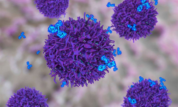
A new high-throughput approach has shown how patients whose tumours express CD58 are more likely to respond to CAR T-cell therapy.
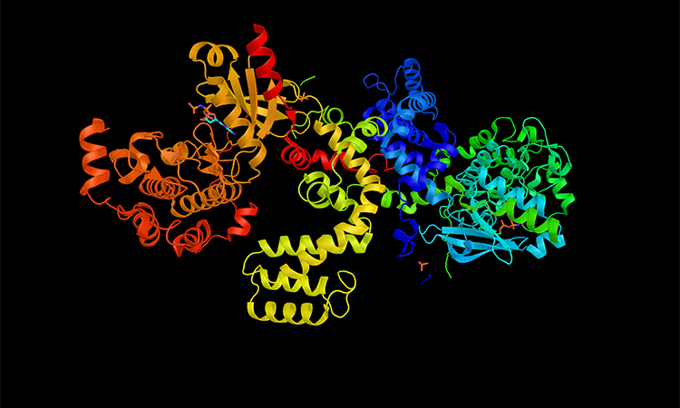
Researchers have developed a 3D structure that allows them to see how and where disease mutations on the twinkle protein can lead to mitochondrial diseases.

A powerful tool to study complex phenotypes and cellular interactions. Map spatial interactions twice as fast with this second-generation system.

Researchers have developed a novel microscopy technique to make invisible molecules visible by “de-crowding” to expand a cell or tissue sample before labelling the molecules.
Using cryo-electron microscopy, researchers have captured the structure of a membrane-bound T-cell receptor complex with bound antigen.
Researchers have developed a novel label-free method named tomographic phase microscopy in flow cytometry for measuring intracellular lipid droplets in 3D.
The monoclonal antibody 19A11 binds E-cadherin, a protein that helps cells stick together, especially in epithelial layers that line the skin, the gut and other organs.

Single-domain antibodies (sdAb) are small, stable antibodies derived from camelids with a single monomeric variable domain.

Researchers for the first time have captured images of an autoantibody bound to a nerve cell surface receptor, revealing the physical mechanism behind a neurological autoimmune disease.
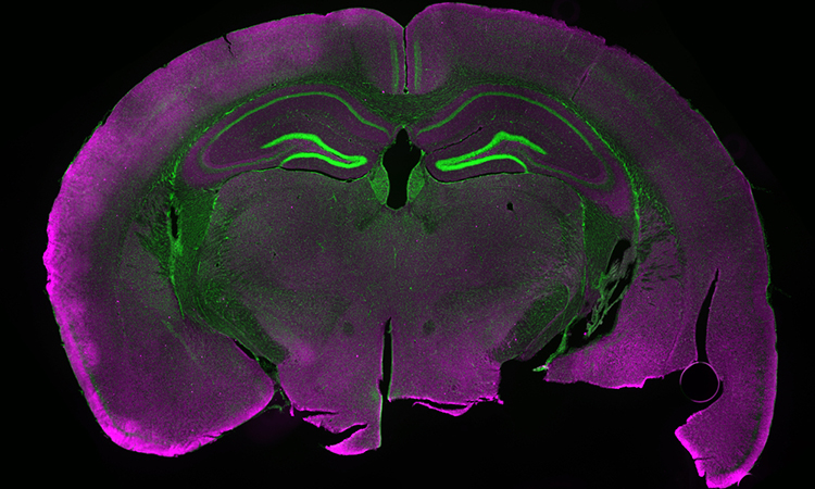
Scientists have developed a new imaging technique that allows researchers to see gene expression and mRNA molecules in the brains of live mice in real time.
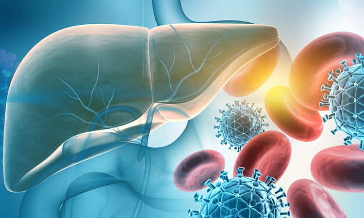
With no current treatments for hepatitis A, scientists have discovered how a protein and enzymes interact to allow hepatitis A virus to replicate.