Cell painting approach used to identify drug compounds with unwanted side effects
Posted: 24 March 2022 | Ria Kakkad (Drug Target Review) | No comments yet
Novel research has shown that morphological fingerprinting could help identify side effects of new bioactive compounds in drug discovery.
A new study, published in Cell Chemical Biology, highlights how researchers from the Max Planck Institute of Molecular Physiology, Germany have developed strategy to reliably detect side effects, such as the disruption of microtubules, at an early stage of searching for bioactive compounds.
According to the researchers, the most used cancer drugs contain active substances that manipulate the cell’s cytoskeleton by binding to microtubules. This can disrupt cell division, impair other essential processes and lead to cell death. However, the surface of microtubules has many deep binding pockets that make them particularly susceptible to modulation by various of chemical substances with diverse chemical scaffolds.
In the search for and development of new active substances, the study of known side effects is necessary. Although there are already standardised test procedures and screenings for identifying undesirable side effects, they still hold many disadvantages. For example, they may not cover all targets in cells, correctly reflect the cellular context or identify targets.
To do this, the researchers used the “cell painting” approach. This is where several functional areas of the cell are stained and then examined microscopically for changes after adding chemical substances. This enables recording hundreds of cellular parameters in a single morphological fingerprint — for example, if one detects similarity of this fingerprint to those of known reference substances, conclusions about the effect of the unknown substance can be drawn.
The value of this approach lies in the possibility of creating fingerprints for thousands of substances in a high-throughput process. The researchers revealed that more than one percent of about 15,000 studied substances had a tubulin-modulating effect. There were also many known reference substances for which an influence on tubulin was previously unknown.
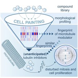
Process of Morphological Profiling
[Credit: @MPI OF MOLECULAR PHYSIOLOGY]
“Reference substances play an essential role in the interpretation of a screen, so they should be carefully evaluated and tested. The compounds identified by the cell painting show a wide variety of chemical scaffolds and even small chemical modifications can have a dramatic impact on the tubulin-binding properties of a compound. This risk is ubiquitous, especially during the compound optimisation phase, where existing atoms are exchanged or removed and new atoms are added in order to improve the pharmacological properties. Additional morphological profiling during the search for hits and their optimisation could not only help unmask side effects such as tubulin modulation early on, but also identify desired and new bioactivities,” said Slava Ziegler, lead researcher. “Moreover, this approach could save time and money as it helps to early assess whether a promising substance has what it takes to become a useful compound or not.”
Related topics
Drug Development, Drug Discovery, Drug Discovery Processes, Drug Leads, Drug Targets, Hit-to-Lead, Medicinal Chemistry, Research & Development, Screening, Technology
Related organisations
Max Planck Institute for Molecular Physiology
Related people
Slava Ziegler