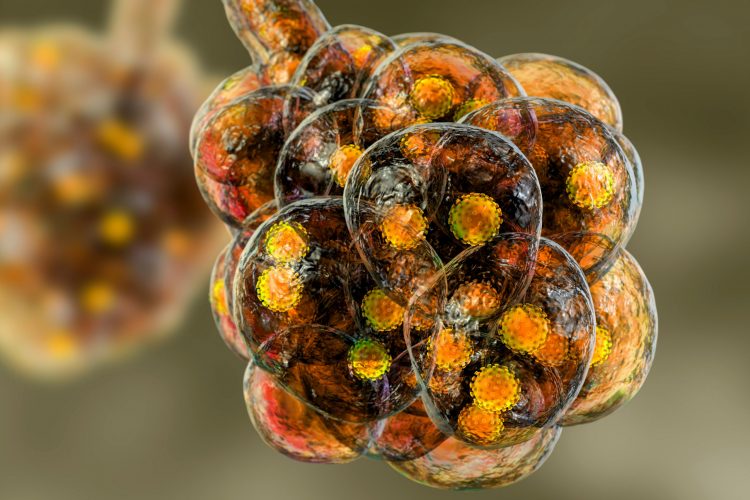
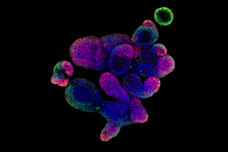
Researchers develop computer algorithm to identify cancer cell lines
Scientists have trained a computer algorithm to identify differences in cancer cell lines based on microscopic images, preventing the misclassification of cells.
List view / Grid view
Scientists have trained a computer algorithm to identify differences in cancer cell lines based on microscopic images, preventing the misclassification of cells.
A new subspace mass spectrometry imaging method has enabled researchers to provide data sets exponentially faster.
This article provides a brief overview of the technical and conceptual advantages of Raman spectroscopy, a label-free imaging technique that is being increasingly used for the purpose of drug evaluation.
A new study has identified the mechanisms through which the SARS-CoV-2 virus enters the brain and how the immune system responds once it does.
A new imaging method called FLASH can provide a visualisation of several tissue types in a 3D format, its developers say.
In this journal, find articles discussing antimicrobial resistance, exploring why inhibiting the interaction between SARS-CoV-2 and neuropilin-1 could help combat COVID-19, as well as how CRISPR can be used to enhance productivity in cell line development. Also in this issue, features on engineering new biologic drugs and precision medicine.
The study shows how drug-like small molecules inhibit the activity of Transient Receptor Potential Canonical 1/4/5 (TRPC1/4/5) channels and could transform the development of future therapies.
Scientists have shown how SARS-CoV-2 induces changes in the architecture of host cells to drive replication and made their data available to all.
Using atomistic simulations, a team has demonstrated how coronavirus Spike proteins move and vibrate to let the virus through cell walls.
Using a new CRISPR-Cas9 tagging strategy, researchers have developed a method that enables the imaging of hundreds of proteins in parallel.
Lan Zhu from Arizona State University explains how cryo-EM methods can be used to obtain structural information on membrane proteins such as GPCRs.
The molecular structure of the SARS-CoV-2 Envelope protein has been identified by researchers using nuclear magnetic resonance.
Three separate studies have identified nanobodies – a miniature form of antibodies found in camelid species – that can bind to the SARS-CoV-2 Spike (S) protein and neutralise the virus in cells.
Using their de novo protein design strategy, researchers engineered human angiotensin converting enzyme 2 (hACE2) protein decoys that can protect cells from SARS-CoV-2 infection.
Scientists reveal that coronaviruses de-activate lysosomes before using them to exit infected cells and spread through the body.