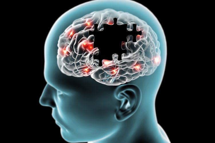
Study identifies pathway responsible for blood-brain barrier impairment in Alzheimer’s
Posted: 9 June 2020 | Hannah Balfour (Drug Target Review) | No comments yet
The team found that the expression of the APOE4 gene variant in pericytes caused amyloid plaque formation in a blood-brain barrier model.
Development of a model of the human blood-brain barrier (BBB) has enabled researchers to discover how APOE4 gene expression causes amyloid plaques to disrupt the brain’s vasculature in Alzheimer’s disease (AD). The team also demonstrated that cyclosporine A and FK506, drugs already approved for use in human, can block the formation of these pathologies.
Around a quarter of people express a variant of the APOE gene, called APOE4, which significantly increases their risk of developing AD. The majority of AD patients, and some elderly people without the disease, suffer from cerebral amyloid angiopathy (CAA). CAA is a condition in which amyloid protein deposits on blood vessel walls impairs the ability of the BBB to properly transport nutrients, clear out waste and prevent the invasion of pathogens and unwanted substances into the brain.
Neuroscientists at MIT’s Picower Institute for Learning and Memory revealed in their study that a vascular cell type, called pericytes, and the calcineurin/NFAT molecular pathway enable the APOE4 variant to promote CAA pathology.
Li-Huei Tsai, study senior author, Picower Professor of Neuroscience and director of the Picower Institute explained that the research indicates that in people with the APOE4 variant, pericytes in their vessels churn out too much APOE protein. APOE causes amyloid proteins, which are more abundant in Alzheimer’s disease, to clump together. Meanwhile, the diseased pericytes’ increased activation of the calcineurin/NFAT molecular pathway appears to encourage the elevated APOE expression.
Establishing the role of APOE4
To study how AD, APOE4 and CAA are linked, the team coaxed human induced pluripotent stem cells to become the three types of cells that make up the BBB: brain endothelial cells, astrocytes and pericytes. Pericytes were modelled by mural cells that they tested extensively to ensure they exhibited pericyte-like properties and gene expression.
After two weeks of growth within a three-dimensional hydrogel scaffold, the BBB model cells assembled into vessels exhibiting properties similar to natural BBBs: low permeability to molecules and expression of the same key genes, proteins and molecular pumps. When immersed in culture media high in amyloid proteins, mimicking conditions in AD brains, the lab-grown BBB models exhibited amyloid accumulation like that of human disease.
Once established, the researchers highlighted how APOE4 causes amyloid accumulation by engineering eight different versions of the BBB model, covering all the possible combinations of the three cell types having either APOE3 (a more typical, healthy variant) or APOE4.
When these models were exposed to amyloid-rich media at one month old, only versions with APOE4 pericyte-like mural cells showed excessive accumulation of amyloid proteins. Replacing APOE4 mural cells with APOE3-carrying ones reduced amyloid deposition. Suggesting pericytes expressing APOE4 are the cause of CAA-like pathology.
This finding was validated when the researchers explored expression in samples of human brain vasculature in the prefrontal cortex and the hippocampus, two regions crucially affected in AD. Consistent with the team’s lab BBB model, people with APOE4 showed higher expression of the gene in the vasculature, and specifically in pericytes, than people with APOE3.
“That is a salient point of this paper,” said Tsai. “It’s really cool because it stresses the cell-type specific function of APOE.”
Potential treatments
To establish why APOE4 was so highly expressed by pericytes, the team identified hundreds of transcription factors which were regulated differently by APOE3 and APOE4 pericyte-like mural cells. Then they scoured that list to see which factors specifically impact APOE expression. One set of factors that were overexpressed by APOE4 pericytes were part of the calcineurin/NFAT pathway. They observed similar upregulation of the pathway in pericytes from human hippocampus samples.
To see if elevated signalling through the calcineurin/NFAT pathway caused amyloid deposition and CAA, the team administered cyclosporine A and FK506 to their BBB model. Both these drugs are designed to reduce the activity of this pathway.
The treated BBB models had reduced APOE expression in their pericyte-like mural cells and therefore less APOE4-mediated amyloid deposits. They also tested the drugs in APOE4-carrying mice and saw that the medicines reduced APOE expression and amyloid build-up.
The team noted that the drugs can have significant side effects, so future studies could identify a more potent small molecule drug that would have less off-target effects.
The paper was published in Nature Medicine.
Related topics
Cell-based assays, Disease Research, Drug Repurposing, Drug Targets, Genomics, Neurosciences, Organoids, Protein Expression, Proteomics, Research & Development, Therapeutics
Related conditions
Alzheimer's disease (AD)
Related organisations
MIT's Picower Institute for Learning and Memory
Related people
Li-Huei Tsai