


news
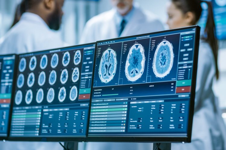
Scientists uncover mechanisms of potential COVID-19 drug targets
Researchers have used CRISPR and cryogenic electron microscopy to unravel the workings of two receptors involved in diseases such as cancer and COVID-19.
List view / Grid view
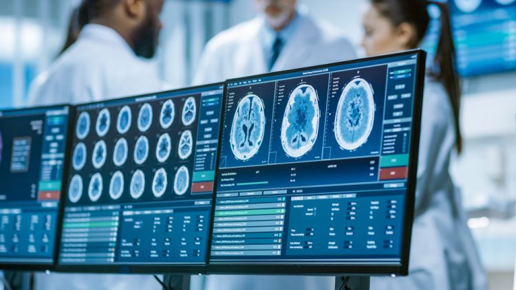
Researchers have used CRISPR and cryogenic electron microscopy to unravel the workings of two receptors involved in diseases such as cancer and COVID-19.
Researchers have developed an inexpensive method for visualising blood flow in the brain that can discern the motions of individual blood cells.
Evaluation of neurotoxicity effects is an active area of investigation in drug discovery and disease modeling.
This whitepaper describes several live-cell phenotypic analyses suitable for the characterisation of astroglia cells.
The cell painting assay uses up to six fluorescent dyes to label and visualize a variety of subcellular structures at the single cell level.

In this original report, find an in-depth analysis of AI and informatics within imaging, synthetic biology, drug screening and drug design. Featured interviews with experts from AstraZeneca, Auransa, PolarisQB and Chalmers University of Technology.
The latest edition of the live-cell analysis handbook is a companion guide for live-cell analysis users. Includes discussion of live-cell analysis.
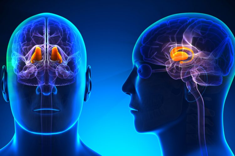
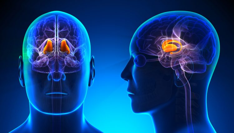
Research shows that cells gather more data inside the thalamus than once believed, potentially changing medicines for brain disorders.
Depletion of ATP due to viral-induced CPE leads to a reduction in luminescence signal, enabling quantitation of viral-induced CPE in host cells.
A non-invasive, label-free optical method can produce high-resolution imaging of cellular brain diseases in vivo.

A US team has designed a high-quality, high-speed imaging system that could lead to new understandings of complex tissue specimens.

10 reasons to choose Bethyl antibodies and reagents from Fortis Life Sciences for your applications.
Scientists have presented a new method for generating the metabolic profiles of cells which could answer questions on conditions such as cancer and liver disease.

Researchers at UT Southwestern have invented a novel microscopy method that could open new avenues of advanced microscopy.

As interest in biotherapeutic proteins grows, the need to reduce cell line development costs and the time to market is more critical than ever.