Application note: In Vivo Imaging with GFP
Posted: 12 November 2019 | Analytik Jena | No comments yet

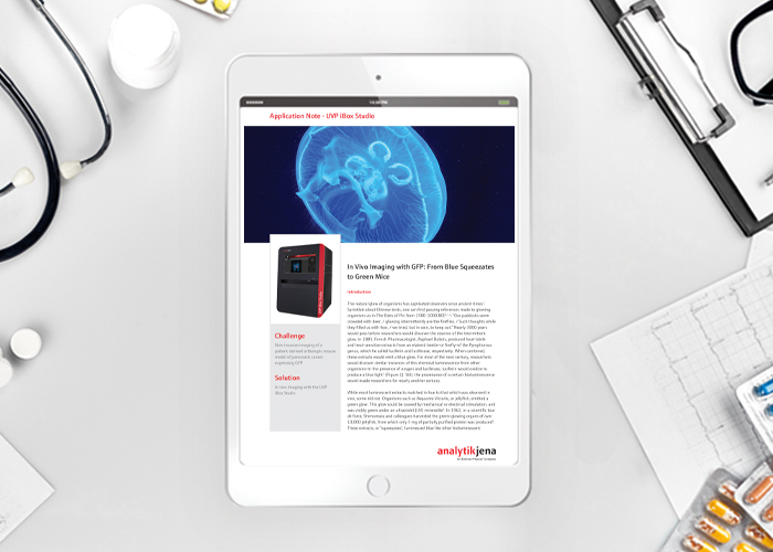
Challenge: Non-invasive imaging of a patient-derived orthotropic mouse model of pancreatic cancer expressing GFP. The solution? In vivo imaging.
In 1885, French Pharmacologist, Raphael Dubois, produced heat-labile and heat-sensitive extracts from an elaterid beetle—or firefly—of the Pyrophorous genus, which he called luciferin and luciferase, respectively. When combined, these extracts would emit a blue glow. For most of the next century, researchers would discover similar instances of this chemical luminescence from other organisms—in the presence of oxygen and luciferase, luciferin would oxidize to produce a blue light. Still, the provenance of a certain bioluminescence would evade researchers for nearly another century.
This Application Note demonstrates the exquisite detail with which the UVP iBox Studio can perform in vivo imaging of an orthotopic mouse model of pancreatic cancer labeled with GFP.
Related content from this organisation
- Unlocking protein potential: liquid handling solutions for drug discovery
- Sample preparation automation in label-free compound profiling
- Scalable solutions for automated sample preparation in label-free compound profiling
- Application note: Automated NGS library preparation on CyBio FeliX
- Application note: qPCR to Measure Minute Volumes in High Throughput
Related topics
Hit-to-Lead, Imaging, In Vivo, Screening, Targets
Related organisations
Analytik Jena