


video
Webinar: Novel flow cytometry ACE-2 blocking assay
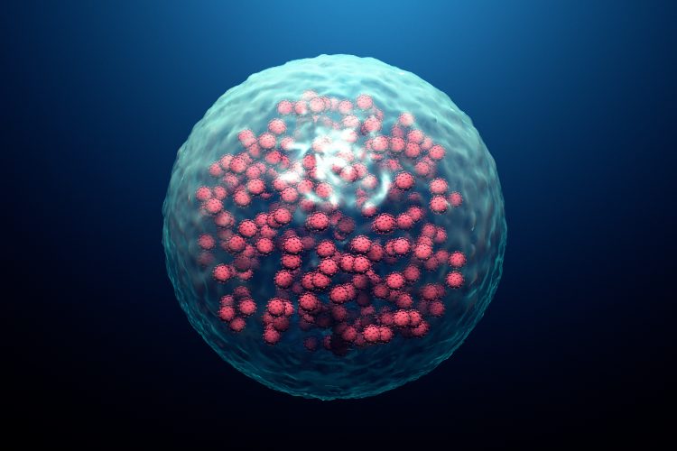
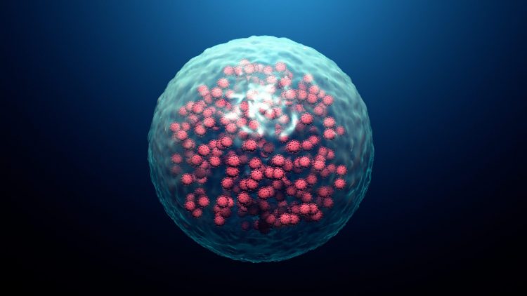
This webinar from Bio-Techne demonstrates how to use a novel in vitro flow cytometry-based assay to monitor SARS-CoV-2 binding to ACE-2.
List view / Grid view
This webinar from Bio-Techne demonstrates how to use a novel in vitro flow cytometry-based assay to monitor SARS-CoV-2 binding to ACE-2.

Discover how workflows are being accelerated to speed up the vaccine research and development process while maintaining safety and immunogenicity.
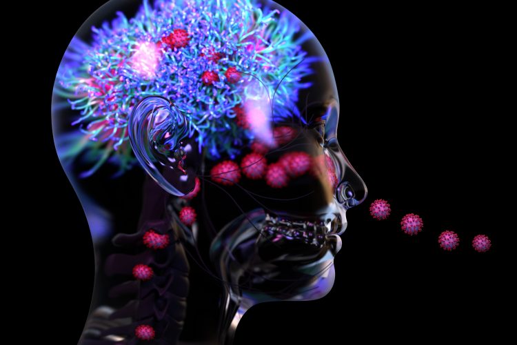
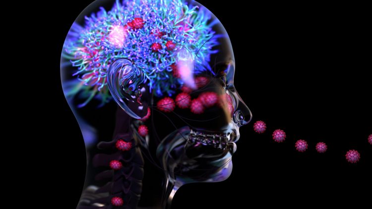
A new study suggests that inflammation and blood vessel damage may be the primary causes of neurological symptoms in COVID-19 patients, instead of the virus infecting the brain.
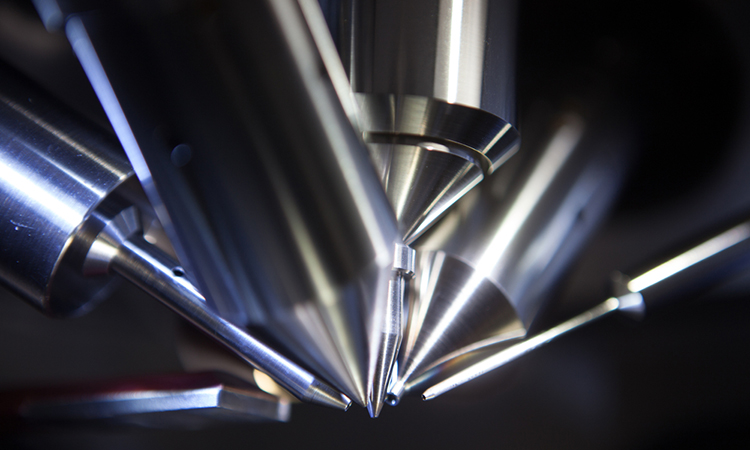
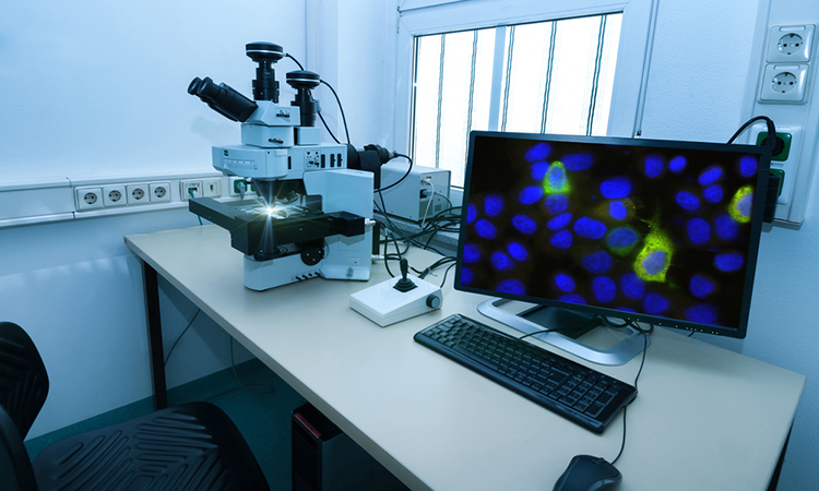
A new subspace mass spectrometry imaging method has enabled researchers to provide data sets exponentially faster.
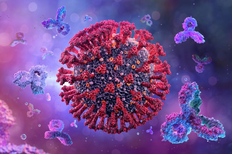
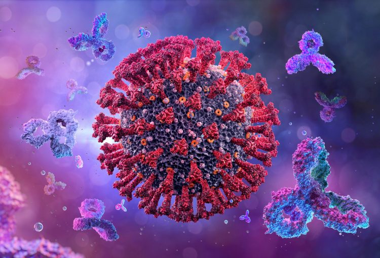
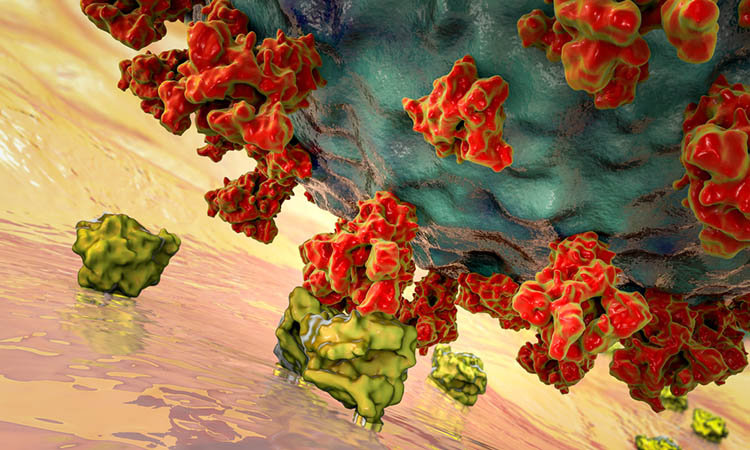
Researchers show that neutralising antibodies targeting the SARS-CoV-2 Spike protein have four distinct structures.

This article provides a brief overview of the technical and conceptual advantages of Raman spectroscopy, a label-free imaging technique that is being increasingly used for the purpose of drug evaluation.
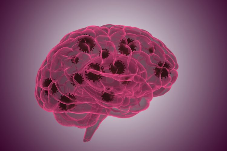
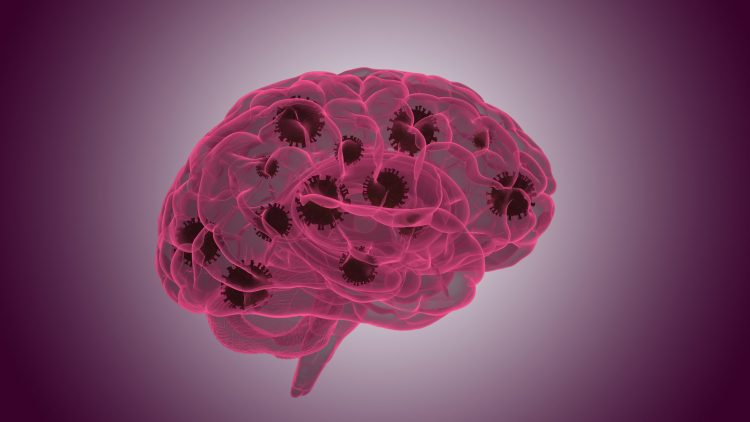
A new study has identified the mechanisms through which the SARS-CoV-2 virus enters the brain and how the immune system responds once it does.
A new imaging method called FLASH can provide a visualisation of several tissue types in a 3D format, its developers say.

In this journal, find articles discussing antimicrobial resistance, exploring why inhibiting the interaction between SARS-CoV-2 and neuropilin-1 could help combat COVID-19, as well as how CRISPR can be used to enhance productivity in cell line development. Also in this issue, features on engineering new biologic drugs and precision medicine.
Scientists have shown how SARS-CoV-2 induces changes in the architecture of host cells to drive replication and made their data available to all.
Using atomistic simulations, a team has demonstrated how coronavirus Spike proteins move and vibrate to let the virus through cell walls.
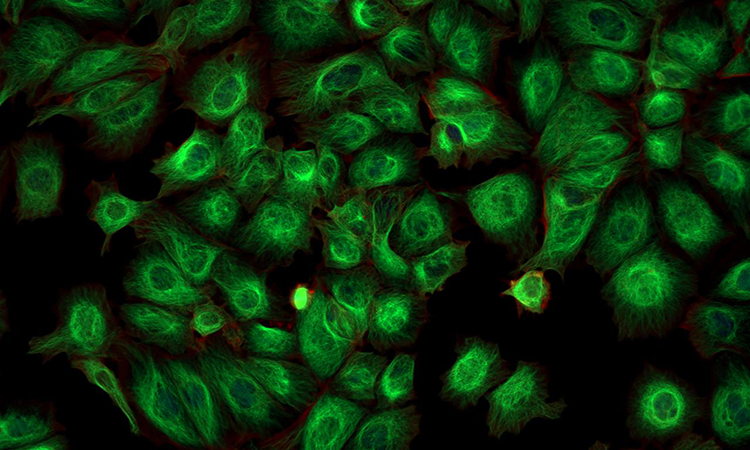
Using a new CRISPR-Cas9 tagging strategy, researchers have developed a method that enables the imaging of hundreds of proteins in parallel.
Lan Zhu from Arizona State University explains how cryo-EM methods can be used to obtain structural information on membrane proteins such as GPCRs.
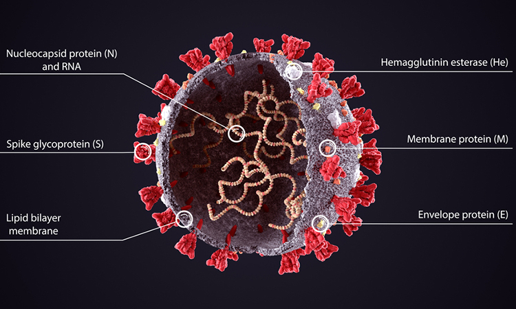
The molecular structure of the SARS-CoV-2 Envelope protein has been identified by researchers using nuclear magnetic resonance.
According to the study, the transcription factor IRF4 drives T cell differentiation and immunosuppression in multiple human cancers.